February 11, 2018 to February 15, 2018
Houston, Texas
By:Bhavesh Ramkorun
SPS Chapter:
I am currently studying physics at a small, but wonderful, liberal arts college, Berea College. We have a small physics program; every year we graduate about three or four students. The physics department currently consist of three faculties who have been constantly encouraging students to participate in summer research projects. Last summer, I had the opportunity to work at the Vanderbilt University Institute of Imaging Science, under Dr. Seth Smith, and Dr. Bryson Reynolds. We also collaborated with Dr. Aashim Bhatia from the Vanderbilt University Children’s Hospital. I used my physics knowledge to read and understand scientific literatures in Diffusion Tensor Imaging, a quantitative imaging technique that an MRI scanner can produce. Our research group processed such images of pediatric population from the children’s hospital. We analyzed our data between normal patients, and patients suffering from pathologies in the spinal cord. By the end of the summer, we had enough data to present at a scientific conference. One of my main objectives at the beginning of last summer was to present at a national conference. With the help of my mentors at Vanderbilt University, I wrote an abstract, and a manuscript, which I submitted, to the international society for optics and photonics (SPIE).
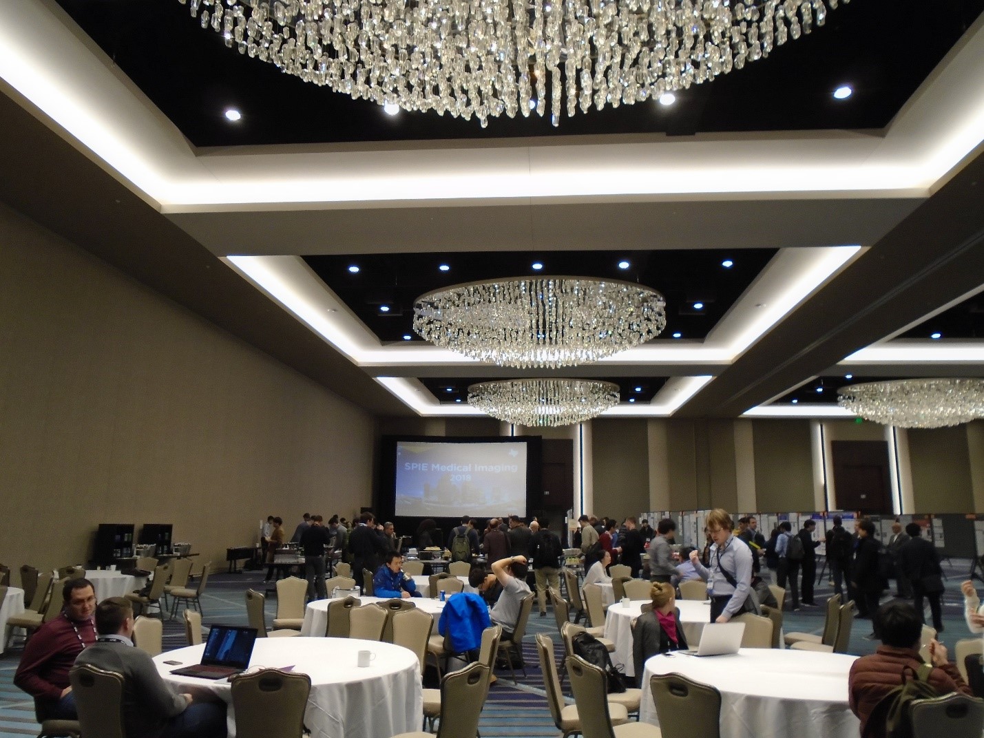
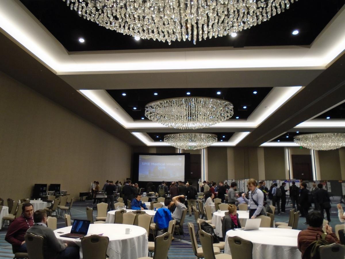
I encountered scientists from around the world doing specialized research in medical imaging. Every room was crowded with scientists (Picture 3). The name tags of attendees indicated if someone was a student; and such, I was able to see many students. All the students, whom I have talked to, were graduate students, in their third or fourth year of their PhD program. When I checked into my hotel, I learned that Houston was expecting 11,000 people for this conference. The conference itself was 5 days long, and I was there for only 3 days. Unfortunately, I did not encounter any other undergraduate students. The Medical Imaging conference itself consisted of nine sub-conferences, details of which are on the SPIE Medical Imaging website (http://spie.org/conferences-and-exhibitions/medical-imaging):
- Physics of Medical Imaging
- Image Processing
- Computer-Aided Diagnosis
- Image-Guided Procedures, Robotic Interventions, and Modeling
- Image Perception, Observer Performance, and Technology Assessment
- Biomedical Applications in Molecular, Structural, and Functional Imaging
- Imaging Informatics for Healthcare, Research, and Applications
- Ultrasonic Imaging and Tomography
- Digital Pathology
There was simultaneous talks of each conference 8 am to 4:30 pm, in different salons, on the fourth floor of the Marriot Marquis Hotel in Houston. In the afternoon, there was poster sessions. I presented my poster on Monday afternoon, in the 6th conference mentioned above, where I talked to faculties, and students who were curious about my project (Picture 1). My presentation lasted 2 hours. During that time, I talked to about 5 faculties, from schools such as Washington University, Vanderbilt University, Bucknell University, as well as faculties from universities around the world, such as Universite de Nantes, and Beijing University. Many of the audience commented that DTI was more famous for brain imaging, than for spinal cord imaging. Therefore, my project was creative, and potentially innovative. Dr Seth Smith and Dr Aashim Bhatia from Vanderbilt University deserve credit for starting this research project.
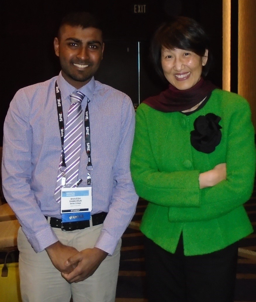
On Tuesday 2/13/2018, the “Physics of Medical Imaging” keynote speaker was Dr. Wei Tse Yang, distinguished chair of diagnostic radiology at MD Anderson Cancer Center. I asked her permission to summarize the physics of her talk in this article. I also took a picture with her (Picture 2). Her research focused on improving diagnosis of breast cancer through quantitative imaging, using at least two novel imaging techniques: 3D optoacoustic imaging (3D OA) and Diffuse Optical Spectroscopic Imaging (DOSI). The breast is imaged with visible light, and near infrared light. In both techniques, light can excite chromophores through either absorption, scattering or reflection, in order to produce topographic maps of the breast. Light reacts with either oxygenated hemoglobin, deoxygenated hemoglobin, lipid or water at different wavelengths to create the topography. 3D OA created accurate 3D maps of the biology of the breast. For example, veins, and arteries were visible at 757 nm, and 1064 nm respectively. 3D OA can also accurately distinguish between the nipple and tumor in the breast, at much better contrast than current popular imaging technique. She then showed an example of how 3D OA detected one inch of breast tumor in a subject; the same tumor was invisible in a mammography. She then summarized data from 3D OA of 2000 patients who demonstrated symptoms of breast cancer. DOSI create 2-D topographic maps of the breast. In one 65-year-old patient, DOSI showed higher water, higher deoxygenated hemoglobin, higher oxygenated hemoglobin, and lower lipid in the breast suffering from tumor, than in the other normal breast. For a 52-year-old patient who was undergoing breast cancer therapy, DOSI data showed a 30% improvement on the tumor, between pre-therapy, and mid-therapy, whereas breast ultrasound data stayed relatively constant. One of Dr. Yang’s conclusion was that 3D OA, and DOSI can reduce inaccurate breast cancer diagnosis, and help minimize invasive surgical intervention.
After the keynote speaker session, I attended a few oral talks of research in physics of medical imaging. Some projects, in the talks that I attended, involved improving photon counting in CT. The talks were theoretical, and assumed knowledge in quantum physics and optics, and many physics that are more advanced. As an undergraduate student with a liberal arts background in Physics, I was unable to follow all the talks, even though I tried to be as attentive as possible. However, the nature of the conference was such that, the audience was specialized, and could easily follow the speaker. Therefore, the speaker could speak about their projects right away, without defining too many terms. After all, they only spoke for about 12 minutes each. I was able to understand the Keynote speaker well, because her research was very similar to my own project at Vanderbilt University.
In the end, I was impressed by the conference in general. I felt welcome, and comfortable interacting with scientists at the conference. I believe that I will have a career in academia as well. The SPIE Medical Imaging is the first of many conferences that I plan to attend in the future.
Scientific Categories: